What is retinopathy of prematurity?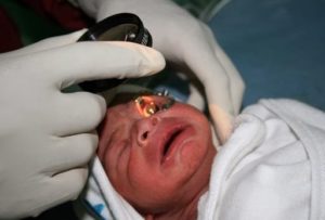
The eye is often compared to a camera. The front of the eye contains a lens that focuses images on the inside of the back of the eye. This area, called the retina, is covered with special nerve cells that react to light.
Underneath the retina is a network of blood vessels. These blood vessels normally grow quickly in the last few weeks before a baby is born. If the baby is born prematurely, there can be a problem with this growth.
In some premature babies, the blood vessels grow into parts of the eye where they do not belong. This can cause scar tissue to form inside the eye. The scar tissue can damage the retina and cause a significant loss of vision. This condition is called retinopathy of prematurity.
Some cases of ROP are mild and correct themselves, but others require surgery to prevent vision loss or blindness. Surgery involves using a laser or other means to stop the growth of the abnormal blood vessels, making sure they don’t pull on the retina.
Because there are varying degrees of ROP, the surgical approach used can differ for each case. The more you know about retinopathy of prematurity and your baby’s surgery, the easier the experience is likely to be for you.
ROP is classified in five stages, ranging from mild (stage I) to severe (stage V):
Stage I — Mildly abnormal blood vessel growth. Many children who develop stage I improve with no treatment and eventually develop normal vision. The disease resolves on its own without further progression.
Stage II — Moderately abnormal blood vessel growth. Many children who develop stage II improve with no treatment and eventually develop normal vision. The disease resolves on its own without further progression.
Stage III — Severely abnormal blood vessel growth. The abnormal blood vessels grow toward the center of the eye instead of following their normal growth pattern along the surface of the retina. Some infants who develop stage III improve with no treatment and eventually develop normal vision. However, when infants have a certain degree of Stage III and “plus disease” develops, treatment is considered. “Plus disease” means that the blood vessels of the retina have become enlarged and twisted, indicating a worsening of the disease. Treatment at this point has a good chance of preventing retinal detachment.
Stage IV — Partially detached retina. Traction from the scar produced by bleeding, abnormal vessels pulls the retina away from the wall of the eye.
Stage V — Completely detached retina and the end stage of the disease. If the eye is left alone at this stage, the baby can have severe visual impairment and even blindness.
Most babies who develop ROP have stages I or II. However, in a small number of babies, ROP worsens, sometimes very rapidly. Untreated ROP threatens to destroy vision.
Causes of ROP
Retinopathy of prematurity causes blood vessels to grow abnormally and randomly in the eye. These abnormal vessels tend to leak or bleed, which leads to scarring of the retina. When the scars shrink, they pull on the retina, causing it to detach from the back of the eye. Since the retina is a vital part of vision, its detachment will cause blindness.
Blood vessels grow from the centre of a developing baby’s retina 16 weeks into the mother’s pregnancy, and then branch outward and reach the edges of the retina 8 months into the pregnancy. In babies born prematurely, normal retinal vessel growth may be disrupted and abnormal vessels can develop, which can cause leaking and bleeding in the eye.
ROP can stop or reverse itself at any point, so it often resolves as the baby grows. Sometimes, though, the disease may progress to cause scarring, which pulls the retina away from the rest of the eye.
ROP has no signs or symptoms. The only way to detect it is through an eye examination by an ophthalmologist.
How is ROP treated?
The most effective proven treatments for ROP are laser therapy or cryotherapy. Laser therapy “burns away” the periphery of the retina, which has no normal blood vessels. With cryotherapy, physicians use an instrument that generates freezing temperatures to briefly touch spots on the surface of the eye that overlie the periphery of the retina. Both laser treatment and cryotherapy destroy the peripheral areas of the retina, slowing or reversing the abnormal growth of blood vessels. Unfortunately, the treatments also destroy some side vision. This is done to save the most important part of our sight the sharp, central vision we need for “straight ahead” activities such as reading, sewing, and driving.
Both laser treatments and cryotherapy are performed only on infants with advanced ROP, particularly stage III with “plus disease.” Both treatments are considered invasive surgeries on the eye, and doctors don’t know the long-term side effects of each.
In the later stages of ROP, other treatment options include:
Scleral buckle: This involves placing a silicone band around the eye and tightening it. This keeps the vitreous gel from pulling on the scar tissue and allows the retina to flatten back down onto the wall of the eye. Infants who have had a sclera buckle need to have the band removed months or years later, since the eye continues to grow; otherwise they will become nearsighted. Sclera buckles are usually performed on infants with stage IV or V.
Vitrectomy: Vitrectomy involves removing the vitreous and replacing it with a saline solution. After the vitreous has been removed, the scar tissue on the retina can be peeled back or cut away, allowing the retina to relax and lay back down against the eye wall. Vitrectomy is performed only at stageV.
What happens if treatment does not work?
While ROP treatment decreases the chances for vision loss, it does not always prevent it. Not all babies respond to ROP treatment, and the disease may get worse. If treatment for ROP does not work, a retinal detachment may develop. Often, only part of the retina detaches (stage IV). When this happens, no further treatments may be needed, since a partial detachment may remain the same or go away without treatment. However, in some instances, physicians may recommend treatment to try to prevent further advancement of the retinal detachment (stage V). If the center of the retina or the entire retina detaches, central vision is threatened, and surgery may be recommended to reattach the retina.
How many infants have retinopathy of prematurity?
Today, with advances in neonatal care, smaller and more premature infants are being saved. These infants are at a much higher risk for ROP. Not all babies who are premature develop ROP. There are approximately 3.9 million infants born in the U.S. each year; of those, about 28,000 weigh 2¾ pounds or less. About 14,000–16,000 of these infants are affected by some degree of ROP. The disease improves and leaves no permanent damage in milder cases of ROP. About 90 percent of all infants with ROP are in the milder category and do not need treatment. However, infants with more severe disease can develop impaired vision or even blindness. About 1,100–1,500 infants annually develop ROP that is severe enough to require medical treatment. About 400–600 infants each year in the US becomes legally blind from ROP.
How successful is treatment for retinopathy of prematurity?
In the small number of infants who need treatment for retinopathy of prematurity, the treatments usually work well at preventing the loss of vision. The most important thing parents can do to help get the best result for their child is to keep all scheduled appointments and follow the doctor’s advice after any treatment.
Retinopathy of prematurity can be very worrisome for parents. There are several groups that offer advice and support for parents, including the Association for Retinopathy of Prematurity and Related Diseases.
Risks and Complications
The goal of surgery for retinopathy of prematurity is to stop the progression of the disease and prevent blindness. Although ROP surgery has a good success rate, not all babies respond to treatment. Up to 25% of babies who have ROP surgery might still lose some or all vision.
Even if the ROP has stopped progressing, vision can still be affected in the following ways:
- strabismus: the eyes aren’t aligned correctly (one example is a “crossed eye”)
- amblyopia: one eye is weaker than the other (sometimes called “lazy eye”)
- myopia: nearsightedness or difficulty seeing distant objects
- glaucoma: increased pressure on the eye that can damage the optic nerve and cause vision loss or blindness
- retinal detachment: the retina detaches from the eye, causing vision loss or blindness
After the Procedure
If admission to the hospital isn’t necessary, you’ll be able to take your child home about an hour after the procedure. Follow-up care for ROP surgery includes giving your child eye drops (to prevent infection) for at least a week.
To make sure the eyes are healing properly and that ROP hasn’t returned, eye exams should be scheduled based on instructions from the ophthalmologist. This is usually every 1-2 weeks. For scleral buckling, the ophthalmologist must examine the buckle every 6 months to account for your child’s growing eye.